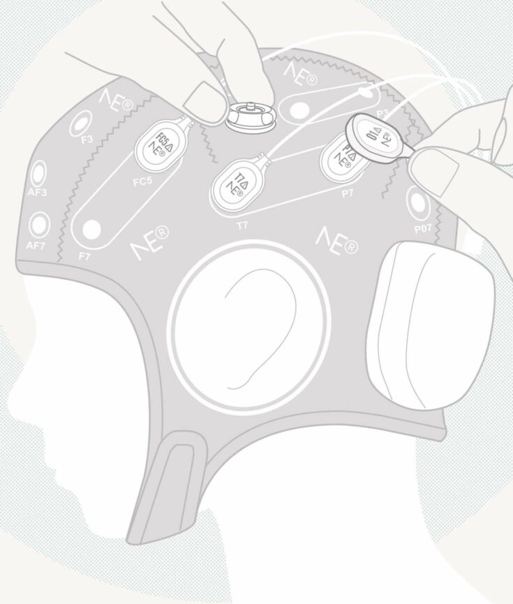
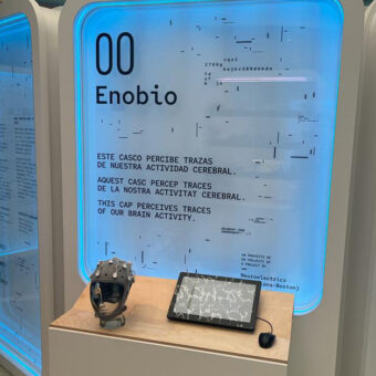
Electroencephalography allows for the recording of neural activity non-invasively by placing a set of electrodes on the scalp. While the fact that electroencephalography is non-invasive has clear advantages for the monitoring of brain activity in humans, it also comes with its limitations.
Neural activity generated by neurons has to propagate through neural tissue, cerebrospinal fluid, skin and bone before reaching the surface electrodes, layers of resistive tissue that alter the original properties of the signal. On top of that, neurons generating the electrical activity are not isolated, but instead, embedded on a complex network of neurons that are constantly active and generating its own electrical activity. All these distortions of the neural signal before it reaches the electrodes placed at the scalp are grouped into the concept of volume conduction effects, technically defined as the distortions an electric (or magnetic) field suffers when it passes through biological tissue towards the measurement sensors. Because of these distortions and diffusions through tissue, scalp sensors can only record brain activity generated at several centimeters below the recording positions. From another prospective, at every position where you put a sensor on the scalp, the recorded brain activity will reflect a weighted sum of the underlying brain sources that span a couple of cm around that position (Makeig et al. 1996).
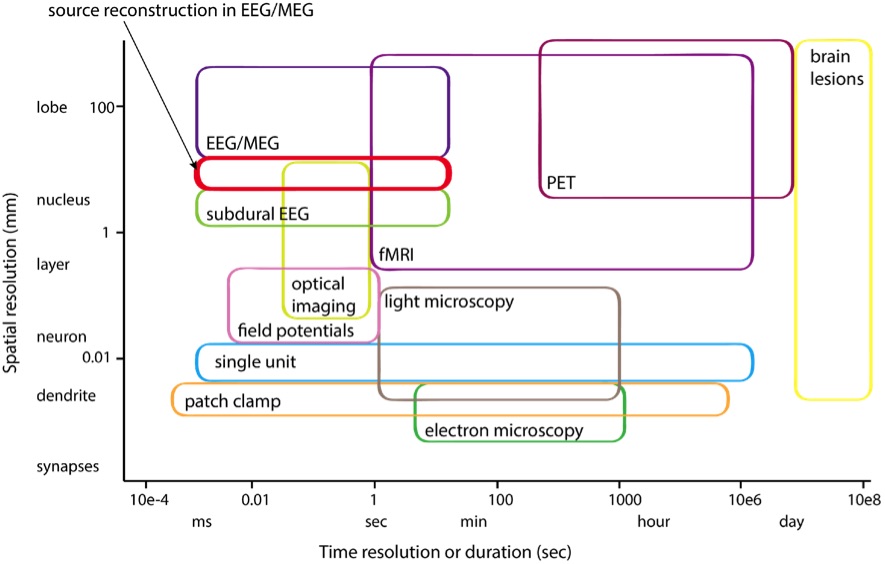
Despite these distortions, EEG remains as one of the most important approaches to study brain activity. While the volume-conduction effect limits us to an estimated spatial resolution of 5-9cm (Nunez 1981), it keeps a temporal resolution on the scale of ms, one of the highest compared to all other methods available for the study of the nervous system (see Figure 1).

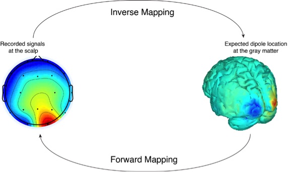
How much can we improve upon these technical limitations? In recent years, there has been remarkable progress in reconstructing the underlying cortical activity from surface electromagnetic data, a set of techniques grouped under the label of source localization or source reconstruction methods (Scherg 1990; Gross et al., 2001; Dale et al. 2000). Intuitively, this technique aims to estimate the spatio-temporal dynamics of currents of the brain that better explain the observed electromagnetic fields through EEG or MEG. In other words, the source localization process consists of calculating, from a set of observed data, what the causal factors that produced them are, what is known as the inverse problem (Figure 2). Note however that, in solving the inverse problem, the number of variables that can be observed (i.e. electrodes we record from) is remarkably small if we compare to the large number of causal factors (i.e. the number of points in the brain where this surface activity could come from, which in practice corresponds to the number of points we would need to create a volume model of a brain). Thus, the inverse problem is, by nature, an ill-posed problem, as we can find more than one solution (brain activity) for an observed scalp voltage.
In order to make this inverse problem well posed, it is necessary to impose additional constraints on the solution. The most common source reconstruction approaches rely on the assumption that sources are temporally uncorrelated, which is particularly appropriate when analyzing the responses sensory stimuli (Mosher et al 1992). Physiological constraints can be introduced by considering that the EEG records signal from populations of neurons localized in the grey matter and are oriented perpendicular to the cortical sheet (Nunez 1981). Knowing the exact shape of the cortical surface through the Magnetic Resonance Imaging (MRI) can further impose anatomical constraints on the head volume conductor model, an approach which is being increasingly used. Constraints on the spatial orientation can also be introduced, especially considering that the non-invasive sensors would record the neural populations that are oriented with a particular angle to the scalp (Miranda et. al 2013). These and other differences would add relative strengths and weaknesses to the different localization methods in terms of accuracy of the approximation, computation time, or spread of the source. For instance, if one expects very few sources, then MAP-estimation may better reflect the true sources while being computationally efficient (Ahlfors et al 1992); if one expects multiple sources with variable spatial extent, then methodologies based on a Sparse-Bayesian Learning may be the most appropriate (Mackay 1992).
While different methods differ on the modeling assumptions, all methods are known to improve spatio-temporal resolution of the EEG, reducing the spatial resolution to 2-4cm (Yao and Dewald 2005; Ding and Lai 2005; Pizzagalli 2007). In the end, the variability in methods to approximate the currents on the brain offer great opportunity to select the most appropriate algorithm for a given experiment.
References:
Ahlfors SP, Ilmoniemi RJ, Hamalainen MS. (1992): Estimates of visually evoked cortical currents. Electroencephalogr Clin Neurophysiol 82(3):225-36
Dale AM, Liu AK, Fischl B, Buckner RL, Belliveau JW, Lewine JD, Halgren E (2000): Dynamic statistical parametric mapping: combining fMRI and MEG to produce high-resolution spatiotemporal maps of cortical activity. Neuron 26:55-67.
Ding, L., Lai, Y., & He, B. (2005). Low resolution brain electromagnetic tomography in a realistic geometry head model: a simulation study. Physics in Medicine and Biology, 50(1), 45.
Gross J, Kujala J, Hamalainen M, Timmermann L, Schnitzler A, Salmelin R. Dynamic imaging of coherent sources: Studying neural interactions in the human brain. Proc Natl Acad Sci USA. 2001 Jan 16;98(2):694-9.
Mackay DJC. (1992): Bayesian Interpolation. Neural Computation 4(3):415-447
Makeig, S., Bell, A., Jung, T.-P., Sejnowski, T., 1996. Independent component analysis of elec- troencephalographic data. In: Touretzky, D., Mozer, M., Hasselmo, M. (Eds.), Advances in Neural Information Processing Systems vol. 8. MIT P, Cambridge MA, pp. 145–151.
Mosher, J.C., Lewis, P.S., and Leahy, R.M. (1992). Multiple dipole modeling and localization from spatio-temporal MEG data. IEEE Trans. Biomed. Eng. 39, 541–557.
Miranda, P. C., Mekonnen, A., Salvador, R., & Ruffini, G. (2013). The electric field in the cortex during transcranial current stimulation. Neuroimage, 70, 48-58.
Nunez, P.L. (1981). Electric Fields of the Brain (New York: Oxford University
Sejnowski, T. J., Churchland, P. S., & Movshon, J. A. (2014). Putting big data to good use in neuroscience. Nature neuroscience, 17(11), 1440-1441.
Scherg M. Fundamentals of dipole source potential analysis. In: Auditory evoked magnetic fields and electric potentials. eds. F. Grandori, M. Hoke and G.L. Romani. Advances in Audiology, vol. 6. Karger, Basel, pp 40-69, 1990.
Pizzagalli, D. A. (2007). Electroencephalography and high-density electrophysiological source localization. Handbook of psychophysiology, 3, 56-84.
Yao, J., & Dewald, J. P. (2005). Evaluation of different cortical source localization methods using simulated and experimental EEG data. Neuroimage,25(2), 369-382.


